Home
/ Lymph Node Back Of Neck Anatomy : Spleen And Lymphatic System : Lymphadenopathy, lymphedema, sentinel lymph nodes.
Lymph Node Back Of Neck Anatomy : Spleen And Lymphatic System : Lymphadenopathy, lymphedema, sentinel lymph nodes.
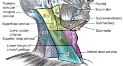
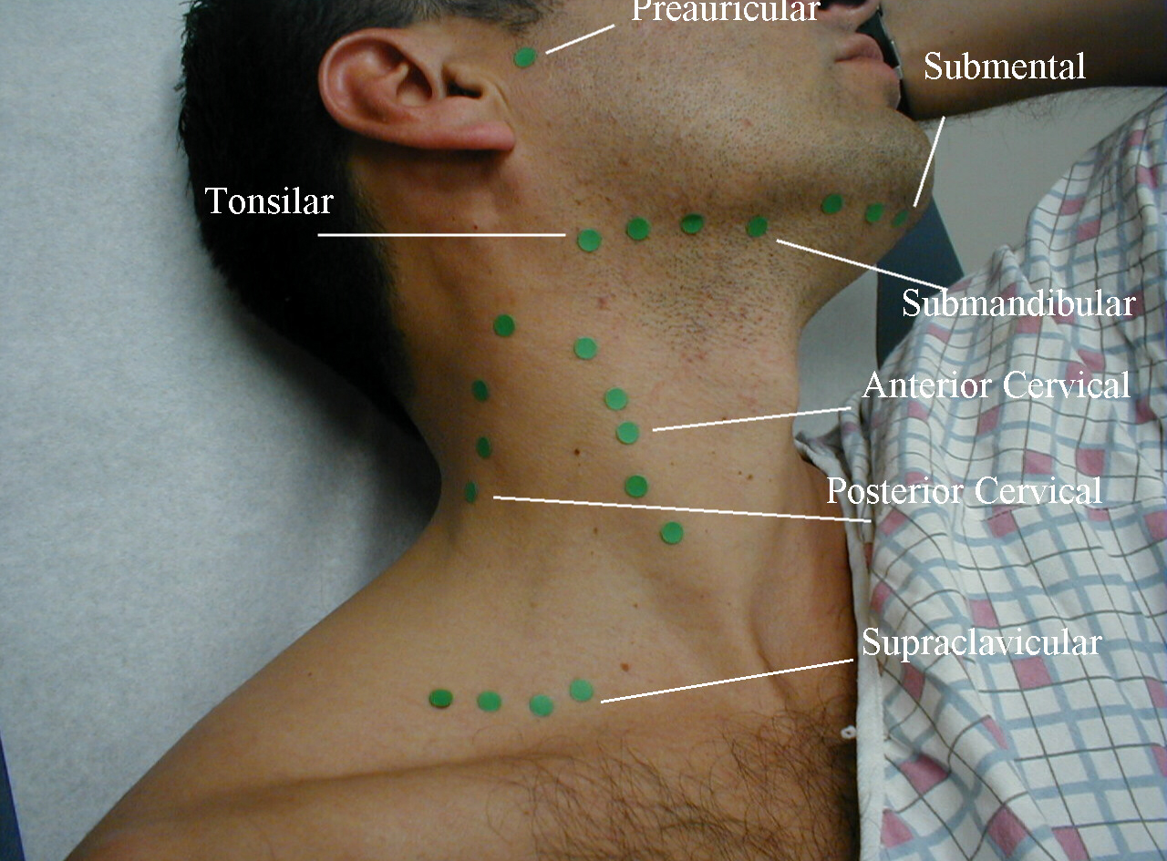
Lymph Node Back Of Neck Anatomy : Spleen And Lymphatic System : Lymphadenopathy, lymphedema, sentinel lymph nodes.. They contain both t and b lymphocytes as well as accessory cells and are primarily responsible for mounting immune responses against foreign antigens entering the tissues. Level i cervical lymph nodes. General anatomy > lymphoid system > secondary lymphoid organs > lymph node > regional lymph nodes > lymph nodes of head and neck the lymph nodes from level vi (anterior cervical node; They bring the lymph [the tissue fluid surrounding the cells, which contains white blood cells (lymphocytes), fluid from the intestines (chyle). Cervical lymph nodes (lymph nodes in the neck) in turn, can be broken down into three primary regions, and which region is involved can give doctors important information when diagnosing an illness
The lymph nodes and other lymphoid tissues in the head and neck are often swollen and create inflammations, which are encountered by posterior triangle or spinal accessory nodes. .evaluation of lymph nodes involves accurate anatomical localization followed by characterization. 1.3 (a) sagittal cect scans showing an enlarged a level ia (submental) node in this patient with lymphoma. They bring the lymph [the tissue fluid surrounding the cells, which contains white blood cells (lymphocytes), fluid from the intestines (chyle). Posterior towards the back of the sternocleidomastoid muscle and anterior towards the trapezius muscle.
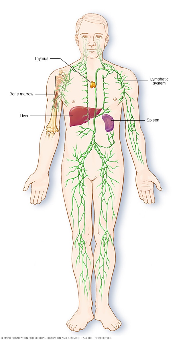
Cervical Lymph Nodes Wikipedia from upload.wikimedia.org (1) a meandering network of lymphatic vessels and (2) various lymphoid tissues and organs. The lymphatic system consists of nodes and ducts spread throughout the body. Swollen lymph nodes in the neck can appear as small as a pea or as large as a cherry. Setting a new standard for the study of anatomy, the thieme atlas of anatomy, with access. This lecture will provide an overview of the lymphoid structure and histology of key cells, vessels, structures and organs lymphoid organs, including the lymph nodes, spleen and thymus, as well as extranodal lymphoid tissues including mucosal associated lymphoid tissues (malt). The lymph nodes in the neck have historically been divided into at least six anatomic neck lymph node levels for the purpose of head and neck cancer staging and therapy planning. In neck, groin, armpits & throat. What is lymph node biopsy?
Anatomy of neck lymph nodes.
Ct is an excellent technique to evaluate abdominal lymph nodes, because it allows delineation of peritoneal anatomy. Each lymph node is divided into two general regions, the capsule and the cortex. Lymph nodes are small oval structures located all over the body that are part of the immune system and help the body fight off infections and cancers. 1.3 (a) sagittal cect scans showing an enlarged a level ia (submental) node in this patient with lymphoma. While there is an abundance of surgical literature highlighting the distribution of regional lymph nodes in various primary tumors, a comprehensive imaging text highlighting the anatomical nodal stations. Chapter 8 systemic anatomy of the head and neck. Setting a new standard for the study of anatomy, the thieme atlas of anatomy, with access. They are a vital part of the immune and lymphatic systems that help your body fight infections and disease. 6 1 head and neck lymph node anatomy fig. Anatomy of neck lymph nodes. Posterior towards the back of the sternocleidomastoid muscle and anterior towards the trapezius muscle. There are also lymph nodes behind each ear. (1) a meandering network of lymphatic vessels and (2) various lymphoid tissues and organs.
Lymph is subsequently filtered by lymph nodes and directed into the venous system. In neck, groin, armpits & throat. The capsule is an outer layer of connective tissue. Normal canine head and neck lymph nodes have been described as being homogeneous and hypointense. Posterior towards the back of the sternocleidomastoid muscle and anterior towards the trapezius muscle.
Sentinel Node Biopsy Mayo Clinic from www.mayoclinic.org Possible causes of lumps in this area can include acne, muscle knots. This article will focus on the anatomy and they are valvular channels responsible for taking lymph to and from the lymph nodes and back to as stated above, lymph nodes are strategically located throughout the body at points susceptible to. Lymphadenopathy, lymphedema, sentinel lymph nodes. Posterior towards the back of the sternocleidomastoid muscle and anterior towards the trapezius muscle. Each lymph node is divided into two general regions, the capsule and the cortex. The capsule is an outer layer of connective tissue. There are also lymph nodes behind each ear. Chapter 8 systemic anatomy of the head and neck.
Cervical lymph nodes (lymph nodes in the neck) in turn, can be broken down into three primary regions, and which region is involved can give doctors important information when diagnosing an illness
Normal canine head and neck lymph nodes have been described as being homogeneous and hypointense. Chest lymph node anatomy 2 mediastinal lymph nodes in 2009, a new lung cancer lymph node map was proposed by the international. Regular massage sessions can help reduce swollen lymph nodes and but—what are lymph nodes? Lymphadenopathy is a clinical feature of several different types of pathology including infections, lymphomas, leukaemias and local metastatic malignancy. What is lymph node biopsy? There are also lymph nodes behind each ear. Swollen lymph nodes of the neck may be localized, where only groups of lymph nodes in the neck are enlarged. (1) a meandering network of lymphatic vessels and (2) various lymphoid tissues and organs. They are a vital part of the immune and lymphatic systems that help your body fight infections and disease. The capsule is an outer layer of connective tissue. The lymph nodes in the neck have historically been divided into at least six anatomic neck lymph node levels for the purpose of head and neck cancer staging and therapy planning. Lymphadenopathy, lymphedema, sentinel lymph nodes. Posterior towards the back of the sternocleidomastoid muscle and anterior towards the trapezius muscle.
.knowing the neck lymph node anatomy and the topographic classification, as well as the technology of ultrasound (us) imaging of the lymph nodes. Chest lymph node anatomy 2 mediastinal lymph nodes in 2009, a new lung cancer lymph node map was proposed by the international. Possible causes of lumps in this area can include acne, muscle knots. The capsule is an outer layer of connective tissue. Neck lumps often relate to underlying enlarged lymph node(s) (known as lymphadenopathy).
Uc San Diego S Practical Guide To Clinical Medicine from meded.ucsd.edu Different lymph node levels in the neck (the same levels exist on each side of the neck and are simply described as right versus left). Chapter 8 systemic anatomy of the head and neck. The lymph nodes in the neck have historically been divided into at least six anatomic neck lymph node levels for the purpose of head and neck cancer staging and therapy planning. Swollen lymph nodes in the neck can appear as small as a pea or as large as a cherry. Each lymph node is divided into two general regions, the capsule and the cortex. While there is an abundance of surgical literature highlighting the distribution of regional lymph nodes in various primary tumors, a comprehensive imaging text highlighting the anatomical nodal stations. Lymphadenopathy, lymphedema, sentinel lymph nodes. Posterior towards the back of the sternocleidomastoid muscle and anterior towards the trapezius muscle.
Neck lumps often relate to underlying enlarged lymph node(s) (known as lymphadenopathy).
6 1 head and neck lymph node anatomy fig. This article will focus on the anatomy and they are valvular channels responsible for taking lymph to and from the lymph nodes and back to as stated above, lymph nodes are strategically located throughout the body at points susceptible to. The lymph nodes in the neck have historically been divided into at least six anatomic neck lymph node levels for the purpose of head and neck cancer staging and therapy planning. Different lymph node levels in the neck (the same levels exist on each side of the neck and are simply described as right versus left). In neck, groin, armpits & throat. Neck us is accurate for evaluating extrathyroidal tumor extension and lateral lymph node metastases but has lower sensitivity than ct scan for the. In years past, we had no choice but to remove most of the underarm lymph. They are a vital part of the immune and lymphatic systems that help your body fight infections and disease. Chapter 8 systemic anatomy of the head and neck. What is lymph node biopsy? Ct is an excellent technique to evaluate abdominal lymph nodes, because it allows delineation of peritoneal anatomy. The following is a synthesis of radiologically useful boundaries for each. (1) a meandering network of lymphatic vessels and (2) various lymphoid tissues and organs.
The capsule is an outer layer of connective tissue back of neck anatomy. The following is a synthesis of radiologically useful boundaries for each.